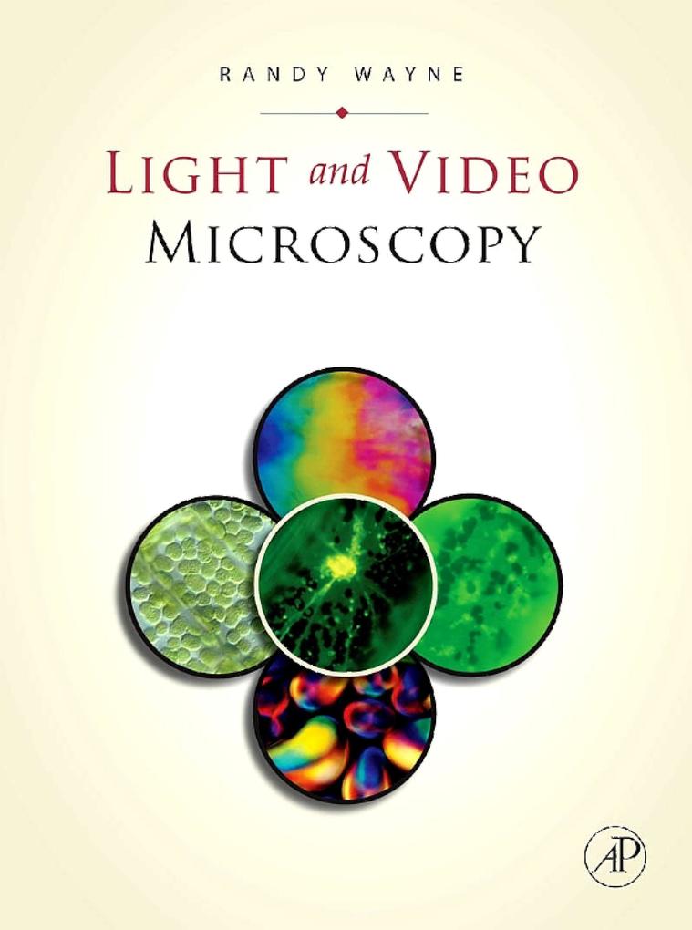
The purpose of this book is to provide the most comprehensive, easy-to-use, and informative guide on light microscopy. Light and Video Microscopy will prepare the reader for the accurate interpretation of an image and understanding of the living cell. With the presentation of geometrical optics, it will assist the reader in understanding image formation and light movement within the microscope. It also provides an explanation of the basic modes of light microscopy and the components of modern electronic imaging systems and guides the reader in determining the physicochemical information of living and developing cells, which influence interpretation.
- Brings together mathematics, physics, and biology to provide a broad and deep understanding of the light microscope
- Clearly develops all ideas from historical and logical foundations
- Laboratory exercises included to assist the reader with practical applications
- Microscope discussions include: bright field microscope, dark field microscope, oblique illumination, phase-contrast microscope, photomicrography, fluorescence microscope, polarization microscope, interference microscope, differential interference microscope, and modulation contrast microscope
Inhaltsverzeichnis
1;Front Cover;1 2;Light and Video Microscopy;4 3;Copyright Page;5 4;Contents;8 5;Preface;12 6;Chapter 1. The Relation between the Object and the Image;14 6.1;Luminous and Nonluminous Objects;14 6.2;Object and Image;14 6.3;Theories of Vision;15 6.4;Light Travels in Straight Lines;18 6.5;Images Formed in a Camera Obscura: Geometric Considerations;18 6.6;Where Does Light Come From?;21 6.7;How Can the Amount of Light Be Measured?;23 7;Chapter 2. The Geometric Relationship between Object and Image;24 7.1;Reflection by a Plane Mirror;24 7.2;Reflection by a Curved Mirror;26 7.3;Reflection from Various Sources;29 7.4;Images Formed by Refraction at a Plane Surface;29 7.5;Images Formed by Refraction at a Curved Surface;33 7.6;Fermat's Principle;39 7.7;Optical Path Length;41 7.8;Lens Aberrations;42 7.9;Geometric Optics and Biology;44 7.10;Geometric Optics of the Human Eye;44 7.11;Web Resources;46 8;Chapter 3. The Dependence of Image Formation on the Nature of Light;48 8.1;Christiaan Huygens and the Invention of the Wave Theory of Light;48 8.2;Thomas Young and the Development of the Wave Theory of Light;52 8.3;James Clerk Maxwell and the Wave Theory of Light;64 8.4;Ernst Abbe and the Relationship of Diffraction to Image Formation;66 8.5;Resolving Power and the Limit of Resolution;73 8.6;Contrast;76 8.7;Web Resource;78 9;Chapter 4. Bright-Field Microscopy;80 9.1;Components of the Microscope;81 9.2;The Optical Paths of the Light Microscope;86 9.3;Using the Bright-Field Microscope;89 9.4;Depth of Field;92 9.5;Out-of-Focus Contrast;93 9.6;Uses of Bright-Field Microscopy;93 9.7;Care and Cleaning of the Light Microscope;93 9.8;Web Resources;94 10;Chapter 5. Photomicrography;96 10.1;Setting up the Microscope for Photomicrography;96 10.2;Scientific History of Photography;98 10.3;General Nature of the Photographic Process;100 10.4;The Resolution of the Film;103 10.5;Exposure and Composition;104 10.6;The Similarities between Film and the Retina;106 10.7;Web Resources;106 11;Chapter 6. Me
thods of Generating Contrast;108 11.1;Dark-Field Microscopy;108 11.2;Rheinberg Illumination;110 11.3;Oblique Illumination;111 11.4;Phase-Contrast Microscopy;112 11.5;Oblique Illumination Reconsidered;121 11.6;Annular Illumination;122 12;Chapter 7. Polarization Microscopy;124 12.1;What Is Polarized Light?;124 12.2;Use an Analyzer to Test for Polarized Light;126 12.3;Production of Polarized Light;127 12.4;Influencing Light;132 12.5;Design of a Polarizing Microscope;133 12.6;What Is the Molecular Basis of Birefringence?;135 12.7;Interference of Polarized Light;139 12.8;The Origin of Colors in Birefringent Specimens;146 12.9;Use of Compensators to Determine the Magnitude and Sign of Birefringence;146 12.10;Crystalline versus Form Birefringence;157 12.11;Orthoscopic versus Conoscopic Observations;158 12.12;Reflected Light Polarization Microscopy;158 12.13;Uses of Polarization Microscopy;158 12.14;Optical Rotatory (or Rotary) Polarization and Optical Rotatory (or Rotary) Dispersion;160 12.15;Web Resources;161 13;Chapter 8. Interference Microscopy;162 13.1;Generation of Interference Colors;162 13.2;The Relationship of Interference Microscopy to Phase-Contrast Microscopy;165 13.3;Quantitative Interference Microscopy: Determination of the Refractive Index, Mass, Concentration of Dry Matter, Concentration of Water, and Density;167 13.4;Source of Errors When Using an Interference Microscope;169 13.5;Making a Coherent Reference Beam;170 13.6;Double-Beam versus Multiple-Beam Interference;171 13.7;Interference Microscopes Based on a Mach-Zehnder Type Interferometer;171 13.8;Interference Microscopes Based on Polarized Light;173 13.9;The Use of Transmission Interference Microscopy in Biology;177 13.10;Reflection-Interference Microscopy;177 13.11;Uses of Reflection-Interference Microscopy in Biology;178 14;Chapter 9. Differential Interference Contrast (DIC) Microscopy;180 14.1;Design of a Transmitted Light Differential Interference Contrast Microscope;180 14.2;Interpretation of a Tr
ansmitted Light Differential Interference Contrast Image;182 14.3;Design of a Reflected Light Differential Interference Contrast Microscope;186 14.4;Interpretation of a Reflected Light Differential Interference Contrast Image;187 15;Chapter 10. Amplitude Modulation Contrast Microscopy;188 15.1;Hoffman Modulation Contrast Microscopy;189 15.2;Reflected Light Hoffman Modulation Contrast Microscopy;192 15.3;The Single-Sideband Edge Enhancement Microscope;192 16;Chapter 11. Fluorescence Microscopy;196 16.1;Discovery of Fluorescence;196 16.2;Physics of Fluorescence;197 16.3;Design of a Fluorescence Microscope;203 16.4;Fluorescence Probes;205 16.5;Pitfalls and Cures in Fluorescence Microscopy;206 16.6;Web Resources;206 17;Chapter 12. Various Types of Microscopes and Accessories;208 17.1;Confocal Microscopes;208 17.2;Laser Microbeam Microscope;211 17.3;Optical Tweezers;212 17.4;Laser Capture Microdissection;212 17.5;Laser Doppler Microscope;213 17.6;Centrifuge Microscope;213 17.7;X-Ray Microscope;214 17.8;Infrared Microscope;215 17.9;Nuclear Magnetic Resonance Imaging Microscope;215 17.10;Stereo Microscopes;215 17.11;Scanning Probe Microscopes;216 17.12;Acoustic Microscope;217 17.13;Horizontal and Traveling Microscopes;218 17.14;Microscopes for Children;218 17.15;Microscope Accessories;218 17.16;Web Resources;219 18;Chapter 13. Video and Digital Microscopy;220 18.1;The Value of Video and Digital Microscopy;220 18.2;Video and Digital Cameras: The Optical to Electrical Signal Converters;221 18.3;Monitors: Conversion of an Electronic Signal into an Optical Signal;229 18.4;Storage of Video and Digital Images;230 18.5;Connecting a Video System;231 18.6;Web Resources;231 19;Chapter 14. Image Processing and Analysis;232 19.1;Analog Image Processing;232 19.2;Digital Image Processing;237 19.3;Enhancement Functions of Digital Image Processors;238 19.4;Analysis Functions of Digital Image Processors;241 19.5;The Ethics of Digital Image Processing;242 19.6;Web Resources;243 20;Chapter 1
5. Laboratory Exercises;244 20.1;Laboratory 1: The Nature of Light and Geometric Optics;244 20.2;Laboratory 2: Physical Optics;246 20.3;Laboratory 3: The Bright-Field Microscope and Image Formation;247 20.4;Laboratory 4: Phase-Contrast Microscopy, Dark-Field Microscopy, Rheinberg Illumination, and Oblique Illumination;249 20.5;Laboratory 5: Fluorescence Microscopy;252 20.6;Laboratory 6: Polarized Light;253 20.7;Laboratory 7: Polarizing Light Microscopy;254 20.8;Laboratory 8: Interference Microscopy;254 20.9;Laboratory 9: Differential Interference Contrast Microscopy and Hoffman Modulation Contrast Microscopy;255 20.10;Laboratory 10: Video and Digital Microscopy and Analog and Digital Image Processing;256 20.11;Commercial Sources for Laboratory Equipment and Specimens;258 21;References;260 22;Appendix I. A Final Exam;286 23;Appendix II. A Microscopist's Model of the Photon;290 24;Index;298 24.1;A;298 24.2;B;298 24.3;C;298 24.4;D;299 24.5;E;299 24.6;F;299 24.7;G;299 24.8;H;300 24.9;I;300 24.10;J;300 24.11;K;300 24.12;L;300 24.13;M;300 24.14;N;301 24.15;O;301 24.16;P;301 24.17;Q;301 24.18;R;301 24.19;S;302 24.20;T;302 24.21;U;302 24.22;V;302 24.23;W;302 24.24;X;302 24.25;Y;302 24.26;Z;302 25;Color Plates;304