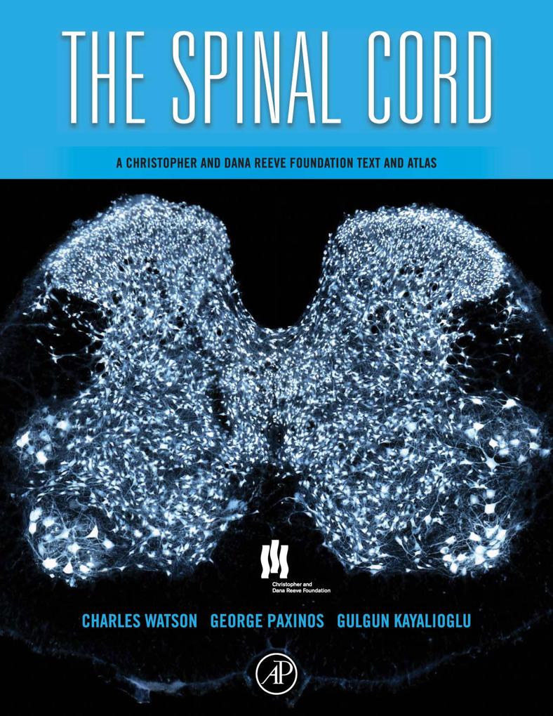
Many hundreds of thousands suffer spinal cord injuries leading to loss of sensation and motor function in the body below the point of injury. Spinal cord research has made some significant strides towards new treatment methods, and is a focus of many laboratories worldwide. In addition, research on the involvement of the spinal cord in pain and the abilities of nervous tissue in the spine to regenerate has increasingly been on the forefront of biomedical research in the past years. The Spinal Cord, a collaboration with the Christopher and Dana Reeve Foundation, is the first comprehensive book on the anatomy of the mammalian spinal cord. Tens of thousands of articles and dozens of books are published on this subject each year, and a great deal of experimental work has been carried out on the rat spinal cord. Despite this, there is no comprehensive and authoritative atlas of the mammalian spinal cord. Almost all of the fine details of spinal cord anatomy must be searched for in journal articles on particular subjects. This book addresses this need by providing both a comprehensive reference on the mammalian spinal cord and a comparative atlas of both rat and mouse spinal cords in one convenient source. The book provides a descriptive survey of the details of mammalian spinal cord anatomy, focusing on the rat with many illustrations from the leading experts in the field and atlases of the rat and the mouse spinal cord. The rat and mouse spinal cord atlas chapters include photographs of Nissl stained transverse sections from each of the spinal cord segments (obtained from a single unfixed spinal cord), detailed diagrams of each of the spinal cord segments pictured, delineating the laminae of Rexed and all other significant neuronal groupings at each level and photographs of additional sections displaying markers such as acetylcholinesterase (AChE), calbindin, calretinin, choline acetlytransferase, neurofilament protein (SMI 32), enkephalin, calcitonin gene-related peptide (CGRP), and neuronal nuclear protein (NeuN).
- The text provides a detailed account of the anatomy of the mammalian spinal cord and surrounding musculoskeletal elements
- The major topics addressed are: development of the spinal cord; the gross anatomy of the spinal cord and its meninges; spinal nerves, nerve roots, and dorsal root ganglia; the vertebral column, vertebral joints, and vertebral muscles; blood supply of the spinal cord; cytoarchitecture and chemoarchitecture of the spinal gray matter; musculotopic anatomy of motoneuron groups; tracts connecting the brain and spinal cord; spinospinal pathways; sympathetic and parasympathetic elements in the spinal cord; neuronal groups and pathways that control micturition; the anatomy of spinal cord injury in experimental animals
- The atlas of the rat and mouse spinal cord has the following features: Photographs of Nissl stained transverse sections from each of 34 spinal segments for the rat and mouse; Detailed diagrams of each of the 34 spinal segments for rat and mouse, delineating the laminae of Rexed and all other significant neuronal groupings at each level. ; Alongside each of the 34 Nissl stained segments, there are additional sections displaying markers such as acetylcholinesterase, calbindin, calretinin, choline acetlytransferase, neurofilament protein (SMI 32), and neuronal nuclear protein (NeuN)
- All the major motoneuron clusters are identified in relation to the individual muscles or muscle groups they supply
Inhaltsverzeichnis
1;Front Cover;1 2;The Spinal Cord: A Christopher and Dana Reeve Foundation Text and Atlas;4 3;Copyright Page;7 4;Contents;10 5;Preface;16 6;Chapter 1 The Organization of the Spinal Cord;18 6.1;Introduction;18 6.2;The gross anatomy of the spinal cord;18 6.3;Spinal cord segments;19 6.4;Spinal nerves;19 6.5;Spinal cord gray and white matter;20 6.6;Lateral cervical nucleus;22 6.7;Lateral spinal nucleus;22 6.8;Onufs nucleus;22 6.9;Central canal;22 6.10;Spinal cord meninges;23 6.11;Vasculature of the spinal cord;23 6.12;References;23 7;Chapter 2 Development of the Spinal Cord;25 7.1;From neural plate to neural tube;25 7.2;Neural crest development;26 7.3;Alar and basal plates and their derivatives;26 7.4;Segmentation of the developing spinal cord;27 7.5;Motoneuron development and cell death;27 7.6;Development of spinal cord afferents and dorsal horn interneurons;29 7.7;Development of glia in the spinal cord;29 7.8;Development of major ascending and descending tracts;29 7.9;Myelination of spinal cord pathways;30 7.10;Relative growth of the spinal cord and vertebral column;30 7.11;References;31 8;Chapter 3 The Vertebral Column and Spinal Meninges;34 8.1;The vertebral column;34 8.2;General features of the vertebrae in mammals;34 8.3;Interspecific variation in vertebral number;36 8.4;The rodent vertebral column;36 8.5;Cervical vertebrae in humans;37 8.6;Thoracic vertebrae in humans;38 8.7;Lumbar vertebrae in humans;38 8.8;The sacrum in humans;38 8.9;The coccyx in humans;39 8.10;Curvatures of the spine;39 8.11;Joints of the vertebrae;40 8.12;Joints between vertebral bodies;40 8.13;Joints between vertebral arches;41 8.14;The craniovertebral joints;41 8.15;Lumbosacral joints;42 8.16;Sacrococcygeal joint;42 8.17;Intercoccygeal joints;43 8.18;Sacro-iliac joints;43 8.19;The intrinsic muscles of the vertebral column;43 8.20;The spinal meninges;45 8.21;Intermediate leptomeningeal layer;47 8.22;References;48 9;Chapter 4 The Spinal Nerves;54 9.1;The anatomy of the dorsal and ventral roo
ts and spinal nerves;54 9.2;Dorsal root (spinal) ganglia;57 9.3;Spinal nerves;59 9.4;Dermatomes;65 9.5;References;67 10;Chapter 5 The Spinal Cord Blood Vessels;74 10.1;Blood flow and spinal cord function;74 10.2;Capillary networks;74 10.3;Spinal cord blood flow imaging;75 10.4;Arterial anatomy;75 10.5;Venous anatomy;77 10.6;Spinal cord lymphatic drainage;77 10.7;Experimental spinal cord ischemia;78 10.8;Blood flow in spinal cord trauma;78 10.9;References;78 11;Chapter 6 Cytoarchitecture of the Spinal Cord;81 11.1;The laminae of Rexed;81 11.2;Lamina 1;81 11.3;Lamina 2;84 11.4;Lamina 3;86 11.5;Lamina 4;87 11.6;The dorsal nucleus;88 11.7;Lamina 5;90 11.8;Lamina 6;91 11.9;Lamina 7;91 11.10;Lamina 8;94 11.11;Lamina 9;95 11.12;Lamina 10;96 11.13;References;97 12;Chapter 7 Localization of Motoneurons in the Spinal Cord;111 12.1;Introduction motoneuron types;111 12.2;Cellular organization of neurons within the ventral and intermediate horns;111 12.3;Experimental approaches to motoneuron localization;112 12.4;Topography of motoneuron pools in the upper cervical spinal cord;113 12.5;Topography of forelimb motoneuron pools in the cervical enlargement;116 12.6;Topography of motoneuron pools in the thoracic and upper lumbar spinal cord;120 12.7;Topography of hindlimb motoneuron pools in the lumbosacral spinal cord;123 12.8;Muscles of the perineum;127 12.9;Deep muscles of the back and tail;127 12.10;Conclusions;128 12.11;References;128 13;Chapter 8 Spinal Autonomic Preganglionic Neurons: the visceral efferent system of the spinal cord;132 13.1;Visceral efferent pathways;132 13.2;Spatial distribution of preganglionic neurons;132 13.3;Morphology of preganglionic neurons and arrangement of their dendrites;133 13.4;Sympathetic preganglionic neurons;134 13.5;Parasympathetic preganglionic neurons;136 13.6;Chemistry of preganglionic neurons;137 13.7;Chemistry of synaptic inputs to preganglionic neurons;137 13.8;Regulation of pelvic organ function;139 13.9;Summary;139 13.10;References;14
0 14;Chapter 9 Central Nervous System Control of Micturition;147 14.1;Introduction;147 14.2;Afferent fibers from bladder to sacral spinal cord;147 14.3;Sensory endings in the bladder wall;147 14.4;Sensory endings in the urethra;148 14.5;Sacral cord;148 14.6;Bladder C-fibers;148 14.7;Bladder A-delta fibers;148 14.8;Ascending projections;149 14.9;Motor innervation of bladder and bladder sphincter;149 14.10;Somatomotor innervation of the external bladder sphincter;151 14.11;Sacral micturition reflexes;153 14.12;Periaqueductal gray;153 14.13;Pontine micturition center (PMC);155 14.14;Continence center or L-region;157 14.15;Other brainstem-spinal pathways possibly involved in bladder and sphincter motoneuronal control;158 14.16;Forebrain micturition control;159 14.17;Conclusion;161 14.18;References;161 15;Chapter 10 Projections from the Spinal Cord to the Brain;165 15.1;Introduction;165 15.2;Ascending spinal projections in the ventrolateral funiculus;165 15.3;Other ascending projections in the ventrolateral funiculus;171 15.4;Projections from the spinal cord to the cerebellum;172 15.5;The dorsal spinocerebellar tract;172 15.6;Dorsal column ascending pathways;174 15.7;References;175 16;Chapter 11 Projections from the Brain to the Spinal Cord;185 16.1;Introduction;185 16.2;The corticospinal tract;185 16.3;Hypothalamic and diencephalic projections to the spinal cord;188 16.4;The rubrospinal tract;188 16.5;The tectospinal tract;189 16.6;Cerebellospinal projections;189 16.7;The reticulospinal tracts;190 16.8;Descending trigeminal and dorsal column nuclei projections;190 16.9;The vestibulospinal tracts;190 16.10;Raphespinal and coeruleospinal tracts;191 16.11;The solitariospinal tract;191 16.12;Projection from the retroambiguus nucleus to the spinal cord;191 16.13;References;191 17;Chapter 12 The Propriospinal System;197 17.1;The propriospinal system: definition and overall function;197 17.2;Subgroups of propriospinal networks;198 17.3;Propriospinal networks and neurotransmitt
ers;200 17.4;Locomotor propriospinal system across species;201 17.5;Propriospinal projections and experimental spinal cord injury;201 17.6;Conclusion;202 17.7;References;203 18;Chapter 13 Spinal Cord Transmitter Substances;208 18.1;Introduction;208 18.2;Cholinergic neurons;208 18.3;Substance P;210 18.4;Noradrenergic projections to the spinal cord;211 18.5;Serotoninergic projections from the raphe nuclei;213 18.6;Dopaminergic projections to the spinal cord;214 18.7;References;215 19;Chapter 14 Spinal Cord Injury: experimental animal models and relation to human therapy;226 19.1;Introduction;226 19.2;General pathophysiology of SCI;226 19.3;Types of spinal cord injury;227 19.4;Immune and inflammatory responses following SCI;228 19.5;Methods to induce spinal cord injury;229 19.6;Assessing functional recovery in animal models of SCI;231 19.7;Assessing human functional recovery;232 19.8;Differences between animal models and humans and functional recovery after SC;233 19.9;Strategies to treat SCI;235 19.10;Clinical trials;239 19.11;References;239 20;Chapter 15 Atlas of the Rat Spinal Cord;255 20.1;Introduction;255 20.2;Methods;255 20.3;Cresyl violet staining and AChE histochemistry;256 20.4;Immunohistochemical processing;256 20.5;Mounting;257 20.6;Photography and diagrams;257 20.7;Nomenclature and abbreviations;257 20.8;Basis of delineation of structures;257 20.9;Naming of spinal cord segments;257 20.10;Identification of regions and segments of the spinal cord in the rat and mouse;258 20.11;References;258 20.12;List of structures;259 20.13;Permission to reproduce figures;260 20.14;Acknowledgements;260 20.15;Mouse spinal cord figures and plates;261 21;Chapter 16 Atlas of the Mouse Spinal Cord;325 21.1;Introduction;325 21.2;Methods;325 21.3;Mouse spinal cord sections provided by the Allen Institute for Brain Science;325 21.4;Photography and diagrams;326 21.5;Basis of delineation of structures;326 21.6;References;326 21.7;List of structures;327 21.8;Permission to reproduce fi
gures;328 21.9;Acknowledgements;328 21.10;Mouse spinal cord figures and plates;329 22;Chapter 17 Toward a Spinal Cord Ontology;397 22.1;What is an ontology?;397 22.2;Regional subdivisions in the spinal cord;397 22.3;A new regional classification based on development;397 22.4;An ontological outline of spinal cord nomenclature;398 22.5;Subdividing the limb enlargements into rostral and caudal groups;398 22.6;Detailed similarities between the arrangement of motoneuron groups in the brachial and lumbar enlargements;398 22.7;Similarities between the segments that immediately precede the upper and lower limb enlargements;399 22.8;Does this spinal cord ontology have any practical application?;400 22.9;References;400 23;Index;401 24;Color Plates Section;405