


Das eBook.de Hörbuch Abo - jederzeit, überall, für nur 7,95 € monatlich!
Jetzt entdecken
mehr erfahren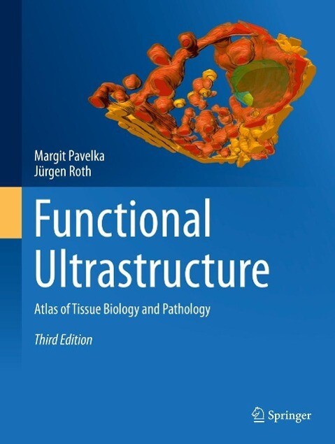
This atlas provides a detailed insight into the complex structure and organization of cells and tissues, and highlights their specific functions as well as the dynamics of diverse intracellular processes. Highly informative electron micrographs are complemented by explanatory texts, selected references and schemes. The concept that subcellular organelles provide the structural foundation for fundamental processes of living organisms is emphasized. The first part covers the cellular organelles and changes caused by experiments or occurring under pathological conditions. The second part employs selected examples to illustrate the principles of functional tissue organization and typical changes resulting from experimental induction or pathological situations. The third edition of the atlas, revised and extended by 23 plates, thus provides an invaluable resource for scientists and students of medicine and biological sciences, particularly of histology, cell and molecular biology. Moreover, it will serve as a handy reference guide for diagnostic and research electron microscopy laboratories in clinical, industrial, and academic settings.
Inhaltsverzeichnis
Introduction. - The Nucleus. - The Cytoplasm: The Secretory System. - The Cytoplasm: The Endocytic System. - The Cytoplasm: Lysosomes and Lysosomal Disorders. - The Cytoplasm: Autophagy. - The Cytoplasm: Mitochondria and Structural Abnormalities. - The Cytoplasm: Peroxisomes and Peroxisomal Diseases. - The Cytoplasm: Paraplasmic Inclusions. - The Cytoplasm: Cytoskeleton. - The Plasma Membrane and Cell Surface Specializations. - Membrane Contact Sites. - Cell-Cell and Cell-Matrix Contacts and Disorders. - Secretory Epithelia. - Resorptive Epithelia. - Sensory Epithelia. - Stratified Epithelia. - Epithelium of the Respiratory Tract. - Urothelium. - Endothelia. - Glomerulus and Disorders. - Connective Tissue. - Adipose Tissue. - Cartilage. - Bone. - Skeletal Muscle and Dystrophies and Myopathy. - Cardiac Muscle. - Smooth Muscle. - Nerve Tissue and Disorders. - Peripheral Blood.
This is a collection of high-quality images
correlated with comprehensive and detailed text to understand the
ultrastructural organization of cells and several tissues. This book is
appropriate for a broad range of audiences with the goal of providing them with
information about the major role that ultrastructural analysis and novel
methods such as FIB-SEM (focused ion beam-scanning electron microcopy) play in
the field of cell and tissue biology and pathology. (Michele Fornaro, Doody s Book Reviews, October, 2015)
Es wurden noch keine Bewertungen abgegeben. Schreiben Sie die erste Bewertung zu "Functional Ultrastructure" und helfen Sie damit anderen bei der Kaufentscheidung.