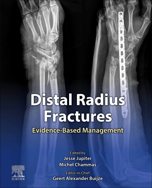
Edited and authored by leading international experts,
Distal Radius Fractures: Evidence-Based Management provides a state-of-the-art overview of diagnosis and management based on today s best practices. Each chapter focuses on one key issue, offering a challenging case and then questioning the reader in an engaging fashion to provide the best available evidence on each topic. Practical and easy to read, this innovative text is a useful resource for all residents, physicians and surgeons who manage fractures.
- Combines current best practices with the knowledge and experience of a global team of expert contributing authors, with a focus on practical use in applying the evidence.
- Covers need-to-know topics such as anatomy and biomechanics, diagnostic management, acute fracture management, management of pediatric and elderly fractures, and more
- Includes valuable case scenarios, technical tips and tricks, and pearls and pitfalls
- Features high-quality illustrations including CT scans, x-rays, and clinical photographs
- Consolidates the latest evidence on distal radius fractures into one convenient resource
- Enhanced eBook version included with purchase, which allows you to access all of the text, figures, and references from the book on a variety of devices
Inhaltsverzeichnis
Section 1. Introduction< BR> 1. Principles of Evidence-Based Management of Distal Radius Fractures< BR> 2. Using Evidence to Manage Distal Radius Fractures< BR> < BR> Section 2. Anatomy and Biomechanics< BR> 3. Epidemiology< BR> 4. Osseous and Ligamentous Anatomy of the Distal Radius< BR> 5. Biomechanics of DRF< BR> < BR> Section 3. Diagnostic Management< BR> 6. Radiographic Parameters and Classifications< BR> 7. Role of Advanced Imaging< BR> < BR> Section 4. Acute Fracture Management< BR> 8. Closed reduction of displaced DRF< BR> 9. Surgical versus conservative interventions for Displaced Distal Radius Fractures< BR> 10. Predictors of Instability and secondary dislocation of conservatively treated DRF< BR> 11. Plating versus Percutaneous Kirschner Wires for simple DRF< BR> 12. Volar plating versus Dorsal plating< BR> 13. Comminuted articular DRF< BR> 14. Comminuted metaphyseal DRF< BR> 15. Bone graft substitutes< BR> 16. Arthroscopic-assisted treatment of DRF< BR> 17. Simultaneous Fractures of the Distal Radius and Scaphoid< BR> 18. Galeazzi Fracture Dislocations< BR> 19. Essex Lopresti Fracture Dislocations< BR> 20. Acute DRUJ Instability< BR> 21. Concomitant ulnar styloid fractures< BR> 22. Persistent Ulnar Sided pain after DRF< BR> 23. Association Between Radiological and Patient-Reported Outcomes< BR> 24. Rehabilitation< BR> 25. Complex Regional Pain Syndrome< BR> < BR> Section 5. Pediatric Fracture Management< BR> 26. Pediatric DRF< BR> 27. Pediatric Forearm Fractures< BR> < BR> Section 6. Elderly Fracture Management< BR> 28. Fracture Fixation in the Elderly< BR> 29. Prevention of DRF< BR> < BR> Section 7. Malunion and Nonunion< BR> 30. Diagnosing the Malunited Distal Radius< BR> 31. Treatment of the Extra-articular Malunited Distal Radius< BR> 32. Treatment of the Intra-articular Malunited Distal Radius< BR> 33. Corrective Osteotomy of Distal Radius Malunion< BR> 34. Three-Dimensional Planning and surgical guidance of Malunion Correction< BR> 35. Nonunion< BR> < BR> Section 8. Posttraumatic Arthritis and Complications< BR> 36. Tendon ruptures after DRF< BR> 37. Chronic DRUJ Instability< BR> 38. Posttraumatic DRUJ Arthritis< BR> 39. Posttraumatic RSL Arthritis< BR> 40. Wrist contracture